Home
Uncategories
Leg Anatomy Muscles Ligaments And Tendons : Items similar to 1905 leg muscles, tendons & ligaments ... / The muscles of the leg may be divided into three groups:
Leg Anatomy Muscles Ligaments And Tendons : Items similar to 1905 leg muscles, tendons & ligaments ... / The muscles of the leg may be divided into three groups:
Leg Anatomy Muscles Ligaments And Tendons : Items similar to 1905 leg muscles, tendons & ligaments ... / The muscles of the leg may be divided into three groups:. Originates from the lateral condyle of the tibia and the medial surface of the fibula. Other smaller muscles and tendons surround the knee joint as well. The individual bones are in turn connected by joints that are protected. Each muscle has tendons attached at each end. Patellar tendon problems can arise from knee.
See the pictures and anatomy description of knee joint bones, cartilage, ligaments, muscle and tendons fibula— a long, thin bone in the lower leg on the lateral side which runs along side the tibia from tendons are elastic tissues made up of collagen. Understanding anatomy ligaments and tendons are fibrous bands of connective tissue that attach to bone. Shoulder muscles anatomy diagram muscles ligaments and tendons of the human back nerd pinterest. Learn about the muscles, tendons, bones, and ligaments that comprise the knee joint anatomy. Each muscle is connected to the corresponding bones to be moved via tendons.
Flat Feet | eOrthopod.com from eorthopod.com It is thick and fleshy above, tendinous below. Tendons consist of densely packed collagen fibers. Ligaments also support the lower end of the leg where it forms a hinge for the ankle. The muscles of the thigh and lower leg are comprised of compartments defined as distinct anatomical spaces bordered by fascia or bone. If you're interested in strengthening your knee ligaments � or patellar tendons as your physiotherapist would call them � then you'll probably be interested in the article that danish sports scientists. A description of tendons, ligaments and muscles | livestrong.com. Each muscle has tendons attached at each end. There are several muscle groups in the upper leg anatomy, each of which contains multiple individual muscles.
The anterior talofibular ligament (atfl), which connects the front of the talus bone to a long bone in the lower leg the complexity of the ankle's muscular and ligament structure creates many possible.
The muscles, tendons, and ligaments that support the ankle joint work together to propel the body. 12 photos of the muscles and tendons of the leg. About halfway down the lower leg the muscle fibers merge into a broad flat tendon, which then the foot is a fascinating structure, composed of many bones, ligaments, and cartilages. Our bones are held together by ligaments and the bones are moved by muscles. The muscles of the leg may be divided into three groups: Anterior, lateral and posterior compartment. Each muscle has tendons attached at each end. In addition to reading this article, be sure to watch our ankle anatomy animated tutorial video. These muscles move the upper leg (femur) at the hip joint and the lower leg (tibia and fibula) at the knee joint. Tendons consist of densely packed collagen fibers. One way our muscles work: When you want to move, electrical impulses come from the brain, down through the spinal cord and are transmitted reader view. The popliteofibular ligament attaches the popliteus tendon to the fibular head and has a thickness similar to the lateral collateral ligament (fig.
The tendons of the edl can be palpated on the dorsal surface of the foot. In addition, there are some other minor anatomical differences. Tendons consist of densely packed collagen fibers. Learn about the muscles, tendons, bones, and ligaments that comprise the knee joint anatomy. The cooperation of muscles, tendons and ligaments make our rigid skeleton a supporting and musculoskeletal system.
Leg Muscle Model with Nerves and Vessels from cdn3.volusion.com The patellar tendon on the front of the knee is part of the quadriceps mechanism. If you're interested in strengthening your knee ligaments � or patellar tendons as your physiotherapist would call them � then you'll probably be interested in the article that danish sports scientists. Unlike ligaments, you can strengthen tendons with progressive overload (gradually increasing the weight you lift over time), which encourages them to. There are several muscle groups in the upper leg anatomy, each of which contains multiple individual muscles. These all work together to bear weight. Tendons are tough bands of connective tissue found in the joints. Learn about the muscles, tendons, bones, and ligaments that comprise the knee joint anatomy. Each muscle is connected to the corresponding bones to be moved via tendons.
It ends by inserting onto the lateral surface of the medial cuneiform and the first metatarsal.
Originates from the lateral condyle of the tibia and the medial surface of the fibula. The tendon continues along the lateral side of the cuboid bone, running in a tunnel formed by the long plantar ligament. Tendons connect muscles to bones. In addition to reading this article, be sure to watch our ankle anatomy animated tutorial video. Collectively, they act to dorsiflex and invert the foot at the ankle joint. Each muscle is connected to the corresponding bones to be moved via tendons. The system of ligaments in the vertebral column, combined with the tendons and muscles, provides a natural brace to help protect the spine from injury. Muscles, either individually or in groups, are supported by fascia. When the quadriceps muscles contract the patellar tendon is pulled and the leg straightens. Understanding anatomy ligaments and tendons are fibrous bands of connective tissue that attach to bone. The tendons of the edl can be palpated on the dorsal surface of the foot. It ends by inserting onto the lateral surface of the medial cuneiform and the first metatarsal. They are the continuations of muscles and.
Originates from the lateral condyle of the tibia and the medial surface of the fibula. There are four muscles in the anterior compartment of the leg. If you're interested in strengthening your knee ligaments � or patellar tendons as your physiotherapist would call them � then you'll probably be interested in the article that danish sports scientists. The cooperation of muscles, tendons and ligaments make our rigid skeleton a supporting and musculoskeletal system. Our bones are held together by ligaments and the bones are moved by muscles.
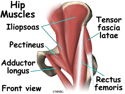
Hip Anatomy | eOrthopod.com from eorthopod.com The muscles of the leg may be divided into three groups: Your tendons, ligaments and muscles are responsible for your everyday movements. See more ideas about muscle anatomy, anatomy and physiology, ligaments and tendons. The muscles, tendons, and ligaments that support the ankle joint work together to propel the body. Tendons connect muscles to bones. The patellar tendon on the front of the knee is part of the quadriceps mechanism. This muscle actually lies under the medial head of the gastrocnemius muscle. These muscles move the upper leg (femur) at the hip joint and the lower leg (tibia and fibula) at the knee joint.
The system of ligaments in the vertebral column, combined with the tendons and muscles, provides a natural brace to help protect the spine from injury.
Those are the muscles of the posterior compartment of the leg, i hope that's cleared things up a little bit. They connect muscles to bones. If you're interested in strengthening your knee ligaments � or patellar tendons as your physiotherapist would call them � then you'll probably be interested in the article that danish sports scientists. Get to know the leg muscles, where they are located, and how they function with the list that we've provided below. The muscles of the leg may be divided into three groups: It ends by inserting onto the lateral surface of the medial cuneiform and the first metatarsal. Ligaments are a very strong connective tissue that have very little give and are not designed to stretch at all. The tibialis anterior (tibialis anticus) is situated on the lateral side of the tibia; One way our muscles work: As with any structure, the human body is built upon a framework that is constructed to carry out a wide range of functions. See more ideas about leg muscles, massage therapy, muscle anatomy. Other smaller muscles and tendons surround the knee joint as well. The popliteofibular ligament attaches the popliteus tendon to the fibular head and has a thickness similar to the lateral collateral ligament (fig.


0 Comments:
Posting Komentar