Home
Uncategories
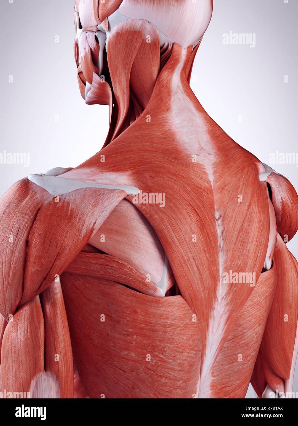
Back Of Neck Anatomy Muscles / Muscles Dos1 120 Ff En Jpg 994 1185 Human Muscle Anatomy Body Anatomy Shoulder Muscle Anatomy - The major muscles of the back, from superficial to deep are divided in three groups:
Back Of Neck Anatomy Muscles / Muscles Dos1 120 Ff En Jpg 994 1185 Human Muscle Anatomy Body Anatomy Shoulder Muscle Anatomy - The major muscles of the back, from superficial to deep are divided in three groups:
Back Of Neck Anatomy Muscles / Muscles Dos1 120 Ff En Jpg 994 1185 Human Muscle Anatomy Body Anatomy Shoulder Muscle Anatomy - The major muscles of the back, from superficial to deep are divided in three groups:. Back muscles are divided into two specific groups: Here the extrinsic back muscles are classified into logical subgroups to facilitate knowledge. Spinous processes of txi to liii and supraspinous ligaments. The head rests on the top part of the vertebral column, with the skull joining at c1. Digastric, mylohyoid, geniohyoid, stylohyoid infrahyoid muscles:
Posterior and lateral views of the neck by phil schatz. This article looks at the anatomy of the back, including bones, muscles, and nerves. Back muscles are divided into two specific groups: The major muscles of the back, from superficial to deep are divided in three groups: Here the extrinsic back muscles are classified into logical subgroups to facilitate knowledge.
Upper Back Pain What S Causing The Top Of My Spine To Hurt from cdn.shopify.com The muscles of the back that work together to support the spine, help keep the body upright and allow twist and bend in many directions. The posterior muscles of the neck are primarily concerned with head movements, like extension. Intermediate back muscles and c. There are many muscles around the neck that help to support the cervical spine and allow you to move your head in different directions. Is the only cutaneous muscle in human body (under the skin) attachments: The back anatomy includes the latissimus dorsi, trapezius, erector spinae, rhomboid, and the teres major. Cervical spine anatomy is quite complex. This article describes the anatomy of the head and neck of the human body, including the brain, bones, muscles, blood vessels, nerves, glands, nose, mouth, teeth, tongue, and throat.
What innervates all of the intrinsic mu… walsh anatomy chapter 2 superficial muscles of the back and neck.
The posterior muscles of the neck are primarily concerned with head movements, like extension. Rectus capitis posterior major and rectus capitis posterior minor attach the inferior the three scalene muscles are found forming the floor of the posterior triangle. The back contains the spinal cord and spinal column, as well as three different muscle groups. The neck muscles, including the sternocleidomastoid and the trapezius, are responsible for the gross motor movement in the muscular system of the head and neck. The back anatomy includes the latissimus dorsi, trapezius, erector spinae, rhomboid, and the teres major. Rectus capitis, longus capitis, longus colli. Adducts, extends and internally rotates the humerus. The pll starts at c2 and goes down the back of the vertebral bodies and intervertebral discs. This article looks at the anatomy of the back, including bones, muscles, and nerves. In anatomy, the neck is also called by its latin names, cervix or collum, although when used alone, in context, the word cervix more often refers to the uterine cervix, the neck of the uterus.3 thus the adjective cervical may refer. Intermediate layer of back muscles. There are several individual muscles within the back anatomy, and it's important to take a quick look the image below to shows all the major back muscles (as well as some neck muscles) Muscles and ligaments work together to support the spine, hold it upright, and control movement during rest and activity.
The muscles of the neck anatomical chart shows in beautiful detail the many anterior, posterior, inferior and lateral views of every muscle that makes up the matrix of support for our skull and brain. The neck muscles, including the sternocleidomastoid and the trapezius, are responsible for the gross motor movement in the muscular system of the head and neck. An interactive tutorial teaching the position, actions, innervation and attachments of the rectus femoris muscle with the aid of anatomical illustrations. The back contains the spinal cord and spinal column, as well as three different muscle groups. The suboccipital muscles act to rotate the head and extend the neck.
Your Back Neck Muscles What They Look Like Bizlinks from bizlinks.files.wordpress.com There are four pairs of muscles that are responsible for chewing movements or mastication. Muscles of the neck are described separately from the compartments. The muscles of the back that work together to support the spine, help keep the body upright and allow twist and bend in many directions. Working in pairs on the left and. There are many muscles around the neck that help to support the cervical spine and allow you to move your head in different directions. This article describes the anatomy of the head and neck of the human body, including the brain, bones, muscles, blood vessels, nerves, glands, nose, mouth, teeth, tongue, and throat. Is the only cutaneous muscle in human body (under the skin) attachments: The splenius muscles originate at the midline and run laterally and.
Rectus capitis, longus capitis, longus colli.
He is the anatomy lead for geeky medics. This article describes the anatomy of the head and neck of the human body, including the brain, bones, muscles, blood vessels, nerves, glands, nose, mouth, teeth, tongue, and throat. Human muscle system, the muscles of the human body that work the skeletal system, that are under voluntary control, and that the following sections provide a basic framework for the understanding of gross human muscular anatomy, with descriptions of the large muscle groups and their actions. Intermediate layer of back muscles. The muscles of the neck run from the base of the skull to the upper back and work together to bend the head and assist in breathing. Is the only cutaneous muscle in human body (under the skin) attachments: Bodies have two kinds of splenius muscles: Neck muscles help support the cervical spine and contribute to movements of the head, neck, upper back, and posterior longitudinal ligament (pll). Brings down corners of the mouth, expressing. The back anatomy includes the latissimus dorsi, trapezius, erector spinae, rhomboid, and the teres major. The muscles of the back that work together to support the spine, help keep the body upright and allow twist and bend in many directions. The major muscles of the back, from superficial to deep are divided in three groups: Inserts on to the humerus.
Spasms of these muscles is a common source of back pain. Digastric, mylohyoid, geniohyoid, stylohyoid infrahyoid muscles: Inserts on to the humerus. Bodies have two kinds of splenius muscles: Extrinsic, intermediate and intrinsic muscles.
Upper Back Muscles High Resolution Stock Photography And Images Alamy from c8.alamy.com Posterior and lateral views of the neck by phil schatz. The back muscles stabilize and move the vertebral column, and are grouped according to the lengths and direction of the fascicles. Along it are easily palpable spinous processes by palpation of the cervical vii and all lying. The major muscles of the back, from superficial to deep are divided in three groups: 21 muscles of the neck: Several other muscles of the back also extend up to the neck region and are partly connected with the cervical part of the vertebral column, including the trapezius, levator scapulae, splenius, iliocostalis, longissimus, rotatores, semispinalis, interspinales, and intertransversarii muscles. This article describes the anatomy of the head and neck of the human body, including the brain, bones, muscles, blood vessels, nerves, glands, nose, mouth, teeth, tongue, and throat. Spasms of these muscles is a common source of back pain.
The back anatomy includes the latissimus dorsi, trapezius, erector spinae, rhomboid, and the teres major.
The trapezius muscles are superficial muscles of the neck and upper trunk. The extensors and rotators of the head and neck: There are four pairs of muscles that are responsible for chewing movements or mastication. Muscles and ligaments work together to support the spine, hold it upright, and control movement during rest and activity. Included are views of the back of the neck, short muscles of the neck, prevertebral muscles. Intermediate layer of back muscles. Back muscles are arranged in several layers, so they are divided into deep and superficial, which, in turn, are arranged in two layers. Neck muscles are bodies of tissue that produce motion in the neck when stimulated. The head rests on the top part of the vertebral column, with the skull joining at c1. By the middle line of the back is a longitudinal groove back (sulcus dorsi). We will attempt to provide a simplified overview of this complex anatomy. Adducts, extends and internally rotates the humerus. He is the anatomy lead for geeky medics.


0 Comments:
Posting Komentar